Parkinson’s disease is a progressive neurological disorder that affects movement, cognition, and overall quality of life. While traditional methods of diagnosing Parkinson’s disease focus on motor symptoms and clinical evaluations, advancements in neuroradiology technology are enhancing the observation of neurological changes. These new methods do not replace a clinical diagnosis but aid in better understanding the condition. Here is more information on some of the most recent advancements in imaging techniques that assist in the exploration of Parkinson’s disease:
Emerging Techniques in Imaging
Newer neuroradiology technologies are pushing the boundaries of how Parkinson’s disease can be explored. Ultrasound imaging of the substantia nigra, also known as transcranial sonography (TCS), is showing promise. This technique enables non-invasive visualization of brain structures and has shown potential in identifying changes associated with Parkinson’s.
High-resolution techniques such as 7-Tesla MRI are also under development. These systems provide a more detailed view of the brain, allowing researchers to study microstructural changes that may previously have gone undetected. Advanced computational analysis integrated with imaging data enables the development of precise models for understanding the disease’s progression.
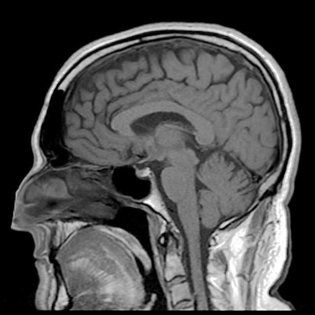
Understanding MRI Scans
Magnetic resonance imaging (MRI) is often used as a tool to study the brain’s structure and function. Although traditional MRI scans may not directly diagnose Parkinson’s, research has identified subtle changes in brain areas affected by the disease. This include areas such as the basal ganglia and substantia nigra.
Diffusion tensor imaging (DTI) is an advanced form of MRI that maps changes in white matter pathways involved in movement. These pathways often show differences in individuals with Parkinson’s compared to those without the condition. Functional MRI (fMRI) is another technique that measures brain activity by detecting blood flow changes, offering insights into how Parkinson’s impacts cognitive and motor functions over time. Together, these imaging technologies provide valuable data for ongoing research.
Utilizing PET and SPECT Scans
Positron emission tomography (PET) and single-photon emission computed tomography (SPECT) scans are imaging methods that measure specific brain functions. These scans have proven particularly useful in studying dopamine, a neurotransmitter that plays a central role in Parkinson’s disease. They provide valuable insights into the progression of the disease and the effectiveness of treatments.
PET and SPECT imaging utilize radiotracers that bind to dopamine transporters in the brain. This helps visualize the reduced dopamine activity characteristic of Parkinson’s. Through these scans, researchers can observe the extent of dopamine loss and explore how it progresses, which may provide valuable information for distinguishing Parkinson’s from other movement disorders with similar symptoms.
Learn More About Neuroradiology
Advancements in imaging have opened new doors for understanding Parkinson’s disease, offering researchers powerful tools to study its effects on the brain. Technologies such as MRI, PET, SPECT, and emerging techniques like transcranial ultrasound and 7-Tesla MRI contribute valuable insights without replacing clinical evaluations. These imaging tools enable more detailed observations of brain structures and functions, enhancing the ability to study disease progression and distinguish Parkinson’s from other conditions. Learn more about the benefits of neuroradiology by consulting with a trusted radiologist.
- Digital Marketing Strategies for UK Businesses: A Complete Guide to Online Growth
- Healthy Living Approaches for Better Work-Life Balance: A Complete Guide to a Happier and Healthier Life
- Modern Urology Solutions for Long-Term Health: A Complete Guide to Better Urinary and Reproductive Wellness
- Preventive Health Management Through Primary Care Options: A Smart Approach to Lifelong Wellness
- Primary Care for Chronic Disease Management: The Key to Better Long-Term Health


Leave a Reply