Heart disease refers to a range of conditions that affect the heart. These conditions can involve blood vessels, heart rhythm, heart muscle, or heart valves. Understanding the structure and function of the heart is fundamental to diagnosing and managing these issues. One of the most effective non-invasive tools available to healthcare professionals for this purpose is an echocardiogram, which provides detailed images of the heart as it works. Here’s more information on diagnosing heart conditions with echocardiograms:
What Is an Echocardiogram?
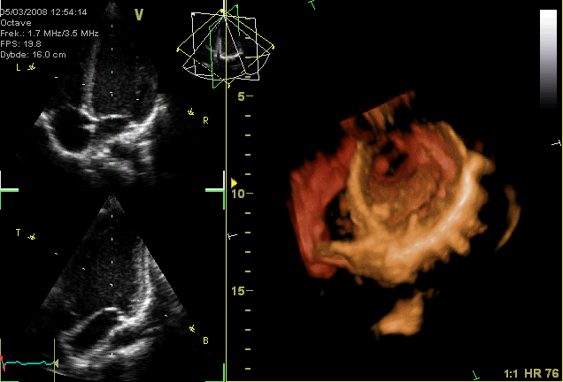
An echocardiogram is a medical test that uses sound waves, or ultrasounds, to create moving pictures of your heart. This procedure allows a specialist to see how your heart is beating and pumping blood. The test is non-invasive and provides information about the size, shape, and function of your heart’s chambers, valves, and surrounding structures.
The Procedure Process
During this diagnostic procedure, a technician applies a gel to your chest and uses a small, handheld device called a transducer. The transducer emits high-frequency sound waves and records the echoes that bounce back from your heart. A computer then converts these echoes into detailed images that are displayed on a monitor.
The Types
There are several types of echocardiograms available, including:
- Transthoracic Echocardiogram (TTE): This is the most common type, where the transducer is placed on the chest wall.
- Transesophageal Echocardiogram (TEE): For more detailed images, a probe is guided down the esophagus to get a closer view of the heart.
- Stress Echocardiogram: This test is performed before and after your heart is stressed, either through exercise or medication, to see how it performs under pressure.
- Doppler Echocardiogram: This technique measures the speed and direction of blood flow within the heart and blood vessels.
How Do Echocardiograms Diagnose Conditions?
Echocardiograms provide real-time, moving pictures of the heart’s structures. This dynamic view is key for diagnostic care, as it allows physicians to assess the heart’s functionality and anatomy. These images reveal the size of the heart chambers, the thickness of the heart walls, and how effectively the heart is contracting and relaxing with each beat.
By observing the heart in motion, specialists can identify abnormalities in the pumping action of its muscle. They also evaluate the function of the heart valves, watching them open and close to verify they are not leaking or narrowed. The moving images help determine if all parts of the heart wall are contributing equally to the heart’s pumping activity. This information is fundamental for identifying areas of damage and for understanding the overall health of the cardiac muscle.
Who Needs an Echocardiogram?
A physician may recommend an echocardiogram for a variety of reasons, often when a patient presents with symptoms like:
- Shortness of Breath
- Chest Pain
- Irregular Heartbeat
It is a valuable tool for investigating the underlying cause of such symptoms. Individuals with certain pre-existing or suspected heart conditions may benefit significantly from this diagnostic test.
An echocardiogram can help in the evaluation of conditions like heart valve disease, where it can detect leaking or stiff valves. It is also used to assess damage to the heart muscle after a heart attack, identify congenital heart defects present from birth, and diagnose conditions like cardiomyopathy. An echocardiogram can also be used to monitor the effectiveness of treatments for various heart conditions.
Schedule an Appointment
If you are experiencing symptoms related to heart conditions, an echocardiogram can provide key information about your heart’s health. This safe and informative procedure enables cardiologists to diagnose and manage heart conditions with precision. Discuss with your healthcare provider whether an echocardiogram is the right next step for you. Schedule a heart consultation today.


Leave a Reply